Lab News

Increasing HDR by putting stem cells back to sleep, published in Cell Reports

When using CRISPR genome editing in stem cells, it’s far easier to break a gene with indels than to fix it with HDR. This manifests in an interesting way. If you...

READ MORE

When using CRISPR genome editing in stem cells, it’s far easier to break a gene with indels than to fix it with HDR. This manifests in an interesting way. If you monitor a “CD34+” population of hematopoietic stem and progenitor cells (HSPCs) from the bone marrow, indels start high and stay high but HDR alleles are lost over time. Why do these different genetic outcomes differ over time? Is HDR bad for the long-term stem cells? Or is editing in the CD34+ population actually heterogeneous, and different cells get different alleles? New work from postdoc Jenny Shin in the lab, out in Cell reports, both answers this question and finds a way to fix the problem.

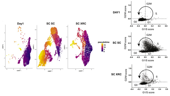
Jenny and collaborators used a powerful combination of immunophenotyping, next generation sequencing, and single-cell RNA-sequencing to investigate and reprogram genome editing outcomes in subpopulations of adult human CD34+ HSPCs. These HSPCs are actually several different types of cells, including more differentiated progenitors that cycle and very “stemmy” long-term HSCs that are quiescent. The team found that there is a dramatic tension between HDR and quiescence in LT-HSCs.  Quiescent stem-enriched cells utilize NHEJ and exhibit almost no HDR. By contrast, non-quiescent cells with the same immunophenotype utilize both NHEJ and HDR. Quiescence is critical for engraftment and stem cell maintenance, so it was now clear that all cells in the CD34+ population get indels and the cycling progenitors were getting HDR alleles, but the quiescent LT-HSCs weren’t doing HDR.

Jenny then had a very creative idea. She asked if a previously reported small molecule cocktail, “XRC”, that maintains quiescence could be used after the fact to re-quiesce LT-HSCs. Using this new strategy and good timing, she found a way to get LT-HSCs with high levels of HDR by briefly allowing them to cycle during editing, and then inducing quiescence later on. This yielded a 6-fold increase in the HDR/NHEJ ratio in quiescent stem cells ex vivo and during long-term engraftment in mouse experiments. The re-quiescence strategy might in future be combined with engineered Cas9-geminin constructs that reduce NHEJ, further tipping the balance towards HDR. Jenny’s results highlight the tradeoffs between editing and fundamental cellular physiology and suggests strategies to manipulate quiescent cells for research and therapeutic genome editing. 
X Close

Welcome to Pia

Pia received her Bachelor’s degree in Biology from ETH Zürich in 2018. She joined the Corn Lab as a Master student in February 2020 working on a CRISPR-based ...

READ MORE

Pia received her Bachelor’s degree in Biology from ETH Zürich in 2018. She joined the Corn Lab as a Master student in February 2020 working on a CRISPR-based screen for regulators in B-cell leukemia. Her research interests include genome engineering, functional genomics, cancer, and DNA repair mechanisms.

X Close

Welcome to Moritz

Moritz joined the Corn lab as a PhD student in July 2020. He has received his Master’s degree in Molecular Biology from the University of Vienna in 2020. Moritz’...

READ MORE

Moritz joined the Corn lab as a PhD student in July 2020. He has received his Master’s degree in Molecular Biology from the University of Vienna in 2020. Moritz’ research interests include functional genomics, therapeutic gene editing and cancer biology, with a special focus on technology development.

X Close

Welcome to Sebastian

Sebastian joined the Corn Lab as a Master student in September 2019 after having received his Bachelor degree in Biological Sciences from the University of...

READ MORE

Sebastian joined the Corn Lab as a Master student in September 2019 after having received his Bachelor degree in Biological Sciences from the University of Konstanz in 2018. He is investigating Fanconi Anemia and the therapeutic potential of CRISPR based systems for this genetic disease.

X Close

Why does fetal globin increase when editing beta globin? Published in Cell Reports

When my lab published editing of the sickle beta globin gene, together with Mark Walters and David Martin, we noticed something quite unexpected. Every time...

READ MORE

When my lab published editing of the sickle beta globin gene, together with Mark Walters and David Martin, we noticed something quite unexpected. Every time we targeted beta globin, we saw fetal globin go up! We also noticed this in beta globin editing papers from the labs of Paula Cannon and Matt Porteus. Transient stress can cause blood cells to express fetal globin, so at first we thought that we were just seeing something short-lived. But it turned out that fetal globin persisted after long-term transplantation of edited human cells into a mouse model. What could be happening in these cells? Could this be a route to treat globin-related diseases such as sickle cell disease? If so, why is beta thalassemia (lack of beta globin) not automatically compensated by fetal globin expression? A new paper from PhD student Mandy Boontanrart, out now in Cell Reports, finally figures out what’s going on. This work was completely driven by Mandy, using a wide range of techniques to dig into a difficult problem.

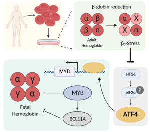
Editing itself can be stressful to cells, but Mandy’s first big findings were that the fetal globin response is not related to the the process of editing. Doing a scan of CRISPR cutting guides through beta globin, she found that any guide targeting an exon causes fetal globin to go up. But equally effective guides that target introns have no effect. She also found that CRISPRi transcriptional repression to knockdown beta globin does increase fetal globin. Therefore, it was something special about lacking beta globin, which is known as B0-stress.

Using clones of blood precursors edited to lack beta globin, Mandy did RNA-seq during differentiation to figure out how cells responded over time. The number one pathway by far was ATF4 signalling. But in a very strange way! It turned out that ATF4 signalling actually went down in cells with beta globin knockout. This was surprising because ATF4 responds to unfolded proteins, it had been suggested that lack of beta globin would lead to alpha chain aggregation that should turn on the unfolded protein response. We were seeing the exact opposite!

This actually makes sense in the context of recent work from the lab of Gerd Blobel, who found out that knockout of the heme-responsive kinase HRI can also increase fetal globin. HRI is upstream of eIF2a and ATF4, so it seemed that free heme (caused by lack of complete adult hemoglobin tetramers) had a stronger role than the free alpha chains in our system.

But what was ATF4 doing to increase fetal globin? Mandy investigated this using a large number of isogenically-controlled ChIP-seq experiments. She found that ATF4 doesn’t bind anywhere in the globin locus, nor does it bind anywhere near BCL11A, which is one of the latest hot globin regulators. In fact, Mandy found that ATF4’s effect on fetal globin still happened in K562 cells, which don’t express any BCL11A at all. Instead, Mandy found strong evidence that ATF4 regulates Myb through binding to a known enhancer region. Myb is itself a regulator of Bcl11A, but can also regulate fetal globin through other factors such as KLF1.

As always there’s a lot more to figure out. And it’s still unclear if we can use these findings to make a difference for patients with globinopathies. But thanks to Mandy’s work, we finally know why fetal globin increases when beta globin is reduced. We also have some clues as to why beta thalassemia is not always compensated by increased fetal globin. In fact, to some extent, it is! Many people with beta thalassemia exhibit increased fetal globin, but not enough to alleviate the symptoms of complete lack of beta globin. ATF4 does many jobs, so it could be that reducing ATF4 enough to yield large amounts of fetal globin might be bad for other biologies. This also adds a note of caution to targeting HRI as a therapeutic angle to increase fetal globin, since doing so could have pleiotropic effects. But all of that remains to be tested!

X Close

Pushing Cas9 off the genome, published in Molecular Cell

Cas9 is a great DNA cutting enzyme, but it’s also a little weird. Unlike other nucleases (such as restriction enzymes), S, pyogenes Cas9 sticks on ...

READ MORE

Cas9 is a great DNA cutting enzyme, but it’s also a little weird. Unlike other nucleases (such as restriction enzymes), S, pyogenes Cas9 sticks on DNA for a looooong time. In fact, it spend the same amount of time on DNA whether it is active or inactive! In a test tube, it takes hours and hours to let go of even a cleaved DNA strand. So how does genome editing even work? Does a human cell care that Cas9 it stuck on its genome? Does it have ways to knock it off?

Superstar grad student Alan Wang’s new paper, out today in Molecular Cell, solves this mystery. Previous work suggested that RNA polymerase II was capable of displacing Cas9 in vitro. But Cas9-based technologies work even when targeted to a non-transcribed region. Alan’s first hint that something interesting was going on came from work in Xenopus egg extract, in collaboration with the lab of Johannes Walter. This “frog juice” is very often used to study DNA repair and can be elegantly deconstructed to figure out biological function. Cas9 in buffer sticks on DNA for a long time, but in Xenopus extract it comes off almost immediately!

Alan took an unbiased approach to figure out what was removing Cas9 from DNA. He fused recombinant Cas9 with a promiscuous biotin ligase, bound purified Cas9-ligase  to a plasmid, and used mass spectrometry to figure out what pushed the Cas9 off the plasmid. We were pleasantly surprised to find that both subunits of a dimeric histone chaperone called FACT were top mass spec hits! Follow-up experiments showed that FACT was necessary and sufficient for displacing Cas9 from DNA substrates. FACT was responsible for turning Cas9 from a multi-turnover “classic” nuclease enzyme into a single-turnover sticky enzyme!

In human cells, FACT had several interesting effects on genome manipulation. Knocking down FACT delayed homology directed repair and altered the balance of repair outcomes. FACT knockdown increased epigenetic marking from both CRISPRi and CRISPRa constructs, and increased CRISPRi phenotypes. We attribute this to increasing the residence time at a target site: giving the effect fused to Cas9 more time to have an effect on the genome. 

The take-home is that cells are not passive players, and play a leading role in genome manipulation. The cell is just as important as the enzyme! Alan’s work starts to reveal how cells monitor their genomes during Cas9 interventions.

X Close

Increasing HDR by timed inhibition of CDC7, published in Nature Communications

When doing genome editing, fixing sequences by HDR is better than breaking them by making indels. If you really want to break something, you could even use HDR...

READ MORE

When doing genome editing, fixing sequences by HDR is better than breaking them by making indels. If you really want to break something, you could even use HDR to insert a precise indel or a stop codon. Unfortunately, HDR is relatively inefficient in human cells. Single stranded oligo donors help, but editing the same locus with a double stranded plasmid DNA donor is almost always painful. What’s the bottleneck? We tried to answer this question in a new paper just out in Nature Communications.

To answer this question, Chris Richardson and Beeke Wienert led a superstar team to perform CRISPRi screening while simultaneously editing with a double stranded DNA plasmid donor. The screen itself was performed by Sharon Feng, a superstar undergraduate. Putting everything together was an exciting collaboration with the labs of Bruce Conklin and Alex Marson.

The first set of hits are known homologous recombination factors, such as BRCA1. This gives high confidence that the screen worked as expected. Surprisingly, the same Fanconi Anemia complexes that are required for single stranded oligo HDR are required for plasmid HDR. The FA pathway is thus a core regulator of all forms of HDR!

But we really wanted to know genes could increase HDR if they were removed. Knocking down a gene is hard to do in many contexts. So we focused on genes with known inhibitors. It turns out that small molecule inhibitors of CDC7 give very nice boosts in HDR from both single stranded oligo and plasmid DNA donors. This works for small changes (SNPs), medium changes (adding epitope tags) and even large cargoes (site-targeted transgenes). It also works in a variety of cell types, including hematopoietic stem cells and T cells. Not every cell is created equally, so check out the paper for detailed guidelines.

Our favorite CDC7 inhibitor is XL413, which is non-toxic and quite reversible. This distinguishes it from some other HDR-improving compounds that lead big genomic messes, including polyploidy. Delving into mechanism, CDC7 inhibition leads to loss of MCM2 phosphorylation. Because MCM2 phosphorylation is required for S phase progression, XL413 leads to a longer S phase. This is a magical phase of the cell cycle for HDR, and so our model is that XL413 increases HDR by increasing the amount of time cells are able to do HDR. We tested this with a timing experiment. Hitting cells with Cas9 and then immediately putting them in XL413 leads to increased HDR, because the cells are piling up in S phase at the same time they are repairing the Cas9 damage. But putting cells in XL413 first and then taking them out during editing leads to decreased HDR. This is because the cells all pile up S phase before editing, and then exit into HDR non-permissive cell cycle while Cas9 is doing its thing.

We hope other labs find XL413 to be useful to increase HDR. It’s not a magic bullet and seems to work especially well in hematopoietic lineages and iPSCs. If you try it out in your favorite cells, please let us know your experience!

X Close

Welcome to Lena

Welcome to Lena Kobel, who joins the lab as a Cell Line Engineer. Lena has a long history in genome engineering, with previous experience in Martin Jinek’s lab...

READ MORE

Welcome to Lena Kobel, who joins the lab as a Cell Line Engineer. Lena has a long history in genome engineering, with previous experience in Martin Jinek’s lab and at Caribou Biosciences. Lena will be working on precision cell models and screens to study the genetics of DNA damage and genome editing.

X Close

ER-autophagy screen published in Cell

Did you know that cells eat their own organelles? This is best known when damaged mitochondria are degraded by autophagy (aka mitophagy). Failure to perform...

READ MORE

Did you know that cells eat their own organelles? This is best known when damaged mitochondria are degraded by autophagy (aka mitophagy). Failure to perform mitophagy can lead to diseases such as Parkinson’s. But many other organelles are also degraded by autophagy. We have been studying autophagy of the endoplasmic reticulum (ER-phagy), which is much less understood than mitophagy. Engulfing a mitochondria in an autophagosome sounds pretty straightforward, but ends up being complicated. Now imagine needing to do that for one part of the ER network! A handful of direct ER-phagy receptors are known. But these receptors are always on the ER, so it was not clear what really initiates and controls ER-phagy. How does the cell know what part to engulf? What are the signals that turn this on and off? What happens when it goes wrong?

Superstars Amos Liang (postdoc) and Emily Lingeman (PhD student) tackled this question in a big way. Using a highly sensitive fluorescent reporter for ER-phagy, they used CRISPRi to ask what genes regulate ER-phagy. The first surprise was that intact mitochondrial oxidative phosphorylation is required to successfully initiate ER-phagy. This is odd because preventing oxidative phosphorylation actually initiates bulk autophagy. But the opposite is true for ER-phagy! The second big surprise was that a weird post-translational modification called UFMylation is required for ER-phagy. Lots of mechanistic work showed how UFMylation machinery is brought to the ER surface and what gets UFMylated during ER-phagy. There are some very interesting parallels to mitophagy, but using totally different machinery. Third, many of the genes involved in ER-phagy are involved in peripheral neuropathy in humans. Since their role in ER-phagy wasn’t previously known, it wasn’t understood how they were connected to cause human disease. This work suggests that failure to do ER-phagy links them all and leads to neurodegeneration. There’s a lot going on here, so read the paper to find out more.  Congrats to Amos and Emily!


X Close

Choosing the right path in genome editing – review published in Nature Cell Biology

Genome editing is all about DNA repair. So if you want to get your cells to do more of what you want (*cough* HDR *cough), you’d better know how they make ...

READ MORE

Genome editing is all about DNA repair. So if you want to get your cells to do more of what you want (*cough* HDR *cough), you’d better know how they make decisions about DNA repair pathways. To help people get the lay of the land, Ph.D. student Charles Yeh and former postdoc Chris Richardson wrote a review all about manipulating DNA repair decisions to influence genome editing outcomes. Out now in Nature Cell Biology!

Be sure to check out Table 1 for a very thorough, non-redundant summary of all the ways people have tried to redirect CRISPR-Cas repair in human cells!


X Close

FILTERS

Please select

Tweets

Contact Us

Questions and/or comments about Corn Lab and its activities may be addressed to:

JACOB.CORN@BIOL.ETHZ.CH